Tag Archives: patient

The Use of Blue Light in Dental Curing

In the dental field, a curing light can use ultraviolet or visible light, depending on what it is designed for. Curing lights are used primarily in the dental industry, where they are used in fillings, sealants, and adhesives for various dental procedures. Other versions of the curing light can be seen in use in manufacturing, where rapid and even curing can be an important part of the manufacturing process. A number of companies produce curing lights which have been custom designed for particular applications, ranging from electronics to dentistry.

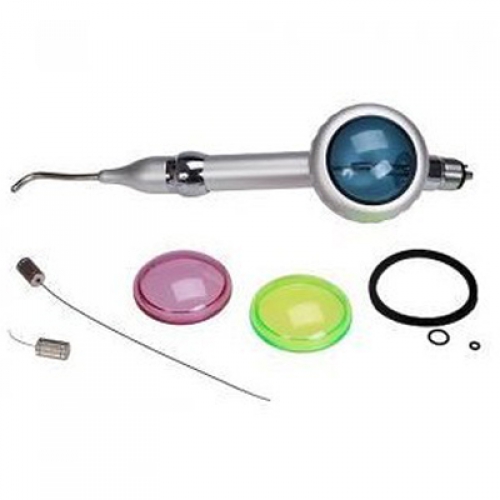
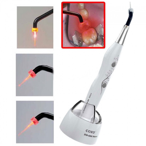
The “blue wand” is a dental curing light. This light is used for polymerization of light-cured resin-based composites or, in other words, the white filling that we put in a tooth. There are several materials that are curable by light. These lights can be Tungsten halogen, LED, plasma, and laser. Halogen and the LED are the most popular.

In the 1960’s the first light-cured resin composite was developed. This led to the first curing light. They called it the”NUVA.” The NUVA used ultraviolet light to cure composites. It was discontinued due to lack of shortened wavelength curing into the resin. Advances were made in the 1980’s in the areas of making visible light curing.This next type of light was the halogen bulb. This light had further penetration and replaced the UV light.

Using a curing light accomplishes two things. In the first place, it makes sure that the resin cures properly and adheres evenly. When applying fillings, this is critical to keep the filling in place in the mouth. For sealants, the curing light limits the risk of cracks and other problems with the sealant. With adhesives for implants and braces, the rapid, even cure is also designed to limit problems in the future.

The dental curing light also increases patient comfort by rapidly curing resins so that the patient is not forced to sit in discomfort while the resin sets. Since the mouth usually needs to be held open wide and may be dry for the procedure, patients usually want the procedure to end as quickly as possible so that they can close their mouths and remoisturize the dried oral membranes. Using a curing light gets patients in and out of the portable folding chair quickly so that the experience of irritation and pain is limited.

The Reason for Using Dental Intraoral Camera

Dentists often find it helpful to be able to show patients exactly what is going on inside their mouths, and to highlight areas where medical attention may be needed. Patients are also less likely to defer or refuse procedures when they can clearly see the area at issue, as some people are suspicious of recommendations for dental procedures, due to concerns about cost, potential pain, or the fears about members of the dental profession.

They say a picture is worth a thousand words. You made the wise decision to purchase an intraoral camera because you thought it would be a good addition to your practice. The problem is that it’s not being used, and currently there is little or no return on your investment.

Offices that use intraoral cameras allow patients to be more interactive in the exam process, which provides patients with a greater sense of understanding and responsibility about personal dental health. Although a traditional visual inspection of the teeth may have sufficed in the past, technology has made it possible for dentists and patients to reap many more benefits from each health exam.

The intraoral camera enlarges the inside of the teeth to more than 40 times their actual size on a full color screen display. By zooming in on problem areas in affecting the teeth, dentists are capable of seeing much more than they could with the human eye alone. Often, dentists find the beginnings of periodontal disease or tooth decay that would have otherwise gone undetected if examined without the intraoral camera.

Images taken by an intraoral camera can also be reviewed later, which can be useful for a dentist who feels a nagging suspicion that something is not quite right in the mouth of a patient. Previously, dentists merely attempted to write an explanation of problems found during exams. Now, dentists can accurately track the progress of treatments or problems for years following a visit.

Furthermore, patients can receive printed pictures of the conditions the dentist finds, which may be beneficial for filing insurance claims. The intraoral camera can also be used to document procedures for legal and educational reasons, and to create projections of a patient’s mouth which can be used in medical schools for the purpose of educating future dentists about various issues which pertain to oral health.

The Reason for Choosing Dental Air Polishing

Air polishing is just that – it’s the practice of polishing the teeth using a stream of air that’s directed onto them. Some air polishing machines may also use a stream of water. The air works in two ways. Firstly, it ‘blows’ onto the teeth and gums to remove any buildups of dirt, and get rid of any food that may have become trapped, which is especially common if you choose not to floss. Secondly, it blows an abrasive powder onto the teeth which helps to tackle stubborn stains such as tea and coffee.

There are two powders that are commonly used for air polishing by dental air polisher, and these are sodium bicarbonate and glycerin powders. These are chosen because of their excellent abrasive qualities. Think about when you’ve got a dirty pan in your kitchen – you may use sodium bicarbonate (baking soda) to remove the stains. It’s exactly the same when it comes to your teeth; sodium bicarbonate can help to get them clean.

A powerful yet controlled jet of water, air and fine powder not only polishes all the surfaces of a tooth, removing plaque, discoloration and soft deposits, but also reaches deep into periodontal pockets up to a depth of 5 mm. It is far more efficient than traditional scrape and polish treatment at removing the damaging biofilm that develops when dental plaque is colonized by bacteria and can cause periodontitis and peri-implantitis to develop. Air flow polishing is completely safe to use with dental implants, veneers, crowns and bridges.

Anyone who has ever cringed as a scraping tool digs into their gums or a polishing disc presses onto tooth enamel will welcome air polishing for its painless, fast and non-invasive method of cleaning. Even deep pockets and interproximal areas are easily reached without uncomfortable and potentially damaging probing by curettes and scrapers and with no instrument contact, the technique does not generate any heat or vibration. The non-toxic powder used in air polishing is also more pleasant and less gritty than the heavy paste used in traditional polishing. Air polishing powders with added flavors, such as spearmint, have even been developed for use in machines, making the experience even more pleasant.

Air polishing has only recently become a common option for dental patients, it’s a concept that’s been around for quite a while. Over the past few years, techniques have been perfected, and air polishing is now believed to be a very safe, effective, and efficient way to remove stains from the teeth, although it’s important to remember that air polishing methods may not be suitable for everyone.

The Research about Dental Air Polisher

Use of dental air polishers decreased as the clinician’s age and experience increased. Curricula in many dental hygiene schools do not include clinical instruction in the use of this polishing device due to inadequate numbers of units and difficulty in moving units between clinic stations. Inadequate or insufficient knowledge and experience, therefore, appears to be a major factor in the underutilization of the air polisher. In an attempt to provide a suitable knowledge base for practicing dental hygienists, the primary purpose of this article is to provide a comprehensive summary and critique of the research on all aspects of air polishing. In addition, a suggested technique, common concerns, and possible solutions will be discussed.

Discussions are based on a review of the relevant literature on air polishing. Tables organize the data into categories to facilitate access of needed information. Because of the various research designs employed and the number of variables that must be controlled, comparative analyses of the studies are difficult. However, where possible, analyses of the validity and reliability of the studies are provided. It should be remembered that while laboratory (in vitro) investigations are useful, the most definitive conclusions must be obtained through clinical (in vivo) studies. Case reports or opinion articles have limited applications. Therefore, interpretation and application of research results must be done with caution.

Air polishing has been compared to scaling and rubber-cup polishing for efficiency and effectiveness of stain and plaque removal. The literature overwhelmingly supports the use of the air polisher as an efficient and effective means of removing extrinsic stain and plaque from tooth surfaces. Air polishing requires less time than traditional polishing methods and removes stain three times as fast as scaling with comers. In addition, less fatigue to the operator has been mentioned as an important benefit of air polishing.

Most investigators agree that intact enamel surfaces are not damaged when stain removal is accomplished with an air polisher. Even after exposure to enamel for the equivalent of a 15-year recall program, surfaces were not altered.

in one in-vitro study, air polishing was shown to remove less root structure than a curet in simulated three-month recalls for three years. Woodall agrees that the air polisher may be preferable to curets in this situation. Since less root structure is removed, decreased root-surface sensitivity also may be a benefit.

Clinical studies to evaluate soft tissue usually provide generalizable conclusions. Gingival bleeding and abrasion are the most common effects of air polishing. These effects are temporary; healing occurs quickly and effects are not clinically significant. No complications were seen with healing at extraction sites following air polishing of teeth prior to extraction. To avoid tissue trauma, the manufacturer recommends pointing the tip of the air polisher at the facial, lingual, or occlusal surfaces, thus avoiding the gingival margins.

Patients also have noticed a salty taste with air polishing, but this was not objectionable. Covering the tongue with moist gauze may prevent irritation and excessive salty taste, as will rinsing with water, mouthwash, or a mint-flavored powder.

What Should You Know about the Dental Air Compressor

Safety should always be your first priority when working with compressed air, no matter the setting. It may seem like a simple tool and you might be tempted to blast a coworker with a burst of air, or use the compressed air to blow dirt or dust away from your skin, but this is dangerous behavior.

Compressed air is under such pressure that even brief contact with protected skin can cause abrasions, cuts and other injuries. If the skin is broken, an air compressor can force air under the skin, causing organ damage or even potentially causing an embolism — an air bubble in the blood that can cause a cardiac event if it reaches the heart. In rare cases, impacts from compressed air have been known to cause traumatic organ damage.

An air compressor could have perfectly functioning filtration and be free of condensate or traveling oil, yet still lag in its performance if air leaks are present at any point between the machine itself and the tools at the end of the pipes.

When air leaks form along a compressed air system, tool performance weakens and operations become less efficient. If an operator is unaware of the problem source, the first solution that often comes to mind is to simply crank up the pressure on the system in order to compensate for the lagging power. This, in turn, leads to increased wear and tear on the internal mechanisms of an air compressor.

Simply put, there’s nothing to be gained from leaks in a compressed air system. The problems associated with leaks are easier to spot and remedy, or prevent altogether, with routine inspections along all the parts that transport air from the compressor to the end tools.

Compressors need lubrication to function, but the oil can get into the airstream, where it could threaten patient health and jeopardize procedures. Some units are oil-free, while others have special sealant systems to prevent leaks. The dental air compressor may also be designed to operate quietly, which can reduce stress for patients who may be worried by the sound of a large engine running near the procedure room.

Buying a name-brand compressor can help reduce that risk even further by giving you access to approved service providers who are trained to maintain your specific piece of equipment. While using a different service provider for your dental equipment maintenance is one option, you aren’t guaranteed to be paired with a technician who knows all the ins and outs of your compressor’s idiosyncrasies.

How to Use Air Polishers Effectively

Use of the dental air polisher for stain removal involves three steps: patient selection and preparation, clinician preparation, and the actual clinical technique. Air polishing should follow a careful review of the patient’s medical and dental history, and a thorough examination of the oral hard and soft tissues. Indications and contraindications, effects on hard tissues, restorations, safety, and alternative uses should be reviewed prior to treatment planning the use of the air polisher.

Preparation of the patient should include an explanation of the procedure, removal of contact lenses, an anti-microbial rinse, application of a lubricant to the lips, placement of safety glasses or a drape over the nose and eyes, and placement of a plastic or disposable drape over the patient’s clothing. Operators should use universal precautions, including protective apparel, a face shield or safety glasses with side shields, gloves, and a well-fitting mask with high-filtration capabilities.

During periodontal surgery, air polishers can prepare root surfaces detoxify them effectively and efficiently, and leave a uniformly smooth root surface that is clean and free of diseased tissues. Dentinal tubules are then occluded, which may result in decreased sensitivity. Superior growth and vitality of human gingival fibroblasts was evident when ultrasonic scaling was followed by air polishing, compared to ultrasonic scaling alone. Air polishing produced root surfaces that were comparable to manually rootplaned surfaces, and provided better access to furcations. Tissue healing following air polishing was comparable to that achieved by hand instrumentation in root preparation during periodontal flap surgery.

Research findings also support the use of air polishing with orthodontic patients. It is the most efficient and effective method for plaque and stain removal around orthodontic brackets, bands, and arch wires. It is not contraindicated on orthodontic bracket composite resin adhesive systems.

In restorative dentistry, air polishers have provided stronger composite repairs than traditional etching gels. They also are superior to rubbercup polishing in preparing occlusal surfaces for etching prior to sealant placement because the rubber cup forces debris into the fissures. Air polishing of occlusal surfaces also allows for deeper penetration of the sealant resin into the enamel surface than rubber cup and pumice cleaning of the fissures. Air polishers also have enhanced sealant bond strength compared to traditional polishing with a low-speed handpiece, bristle brush, pumice, and water.

Oral health care professionals have a responsibility to patients to engage in life-long learning in order to provide the most contemporary clinical care. Air polishing has been studied extensively and, when used appropriately, provides a safe, efficient and contemporary approach to achieving a variety of treatment goals.

The Advantages of Dental Curing Light

In the dental field, a curing light can use ultraviolet or visible light, depending on what it is designed for. Both dentist and patient need to wear eye protection to limit damage to the retina for even the 20 seconds to a minute that the light is in use during rapid curing, and the light needs to be well maintained so that it will work properly and effectively. It’s also important to use the right curing light for the right resin product; many lights are designed to handle a range of resins safety.

Both light intensity – or irradiance – and the dental application should factor into a dentist’s decision regarding his or her choice of curing light. For instance, irradiance is measured by calculating power output, or milliwatts (mW), of a curing light across the surface area of the curing light guide. A curing light must deliver a minimum irradiance of 400mW/cm2 for a time interval to adequately polymerize a 1.5-2mm thick resin composite.

Clinicians also should consider the clinical application at hand. It has been documented that irradiance of curing lights attenuate/decrease significantly when it passes through restorative materials, such as ceramic restorations or resin composites. The percentage of decrease in irradiance depends on filler type, filler loading, shades, refractive index, opacity, translucency and thickness of restorative materials. Curing lights with high irradiance compensate for the decrease in the loss of total energy and allow dentists to cure resin composites completely. In general, an irradiance of 1000mW/cm2 or higher is considered ideal to cure resin-based materials through indirect restorations.

Using a curing light accomplishes two things. In the first place, it makes sure that the resin cures properly and adheres evenly. When applying fillings, this is critical to keep the filling in place in the mouth. For sealants, the curing light limits the risk of cracks and other problems with the sealant. With adhesives for implants and braces, the rapid, even cure is also designed to limit problems in the future.

The dental curing light also increases patient comfort by rapidly curing resins so that the patient is not forced to sit in discomfort while the resin sets. Since the mouth usually needs to be held open wide and may be dry for the procedure, patients usually want the procedure to end as quickly as possible so that they can close their mouths and remoisturize the dried oral membranes. Using a curing light gets patients in and out of the chair quickly so that the experience of irritation and pain is limited.

The Differen Uses of Intraoral Camera

An intraoral camera is a tool your dentist uses to examine your mouth in as detailed a way as possible. The instrument, which may look like an oversized pen, has a camera that takes high-resolution footage or images of a patient’s mouth and shows the visuals real-time on a monitor—they’re like high-tech versions of the hand mirrors you see in your dentist’s practice. There’s much more to intraoral cameras, though.

Cameras can be used to take clear visual records for patient files, and to generate material which can be used in consultations and discussions with other dental providers. For example, a general dentist might use an intraoral camera to take images of a tooth or area of the jaw which requires oral surgery so that a maxillofacial surgeon can examine the information before he or she meets the patient to get an idea of the kind of surgery which might be required.

Each feature that benefits the dentist also benefits the patient—maybe even more. Your dentist understands symptoms and conditions thoroughly, but it’s often difficult to explain precisely what is happening in a patient’s mouth using just a mouth mirror, which is small and hard to see, or an x-ray image, which takes time to print and doesn’t display images clearly.

When your dentist uses an intraoral camera during your examination, however, you’re seeing exactly what he or she sees right then. Dentists can display clear, colorful images, allowing them to point out any issues and discuss them with you immediately. You’ll certainly learn a lot about your mouth! And the more you see and understand, the more confident you can be when making treatment decisions.

The intraoral cameras designed for use in dental facilities come with disposable probes or probe covers to ensure that germs are not passed between patients, and they may come with a variety of options which enhance the functionality of the camera. Versions designed for home use are usually much more basic, but they can still be useful for people who want to see the inside of the mouth. Using a camera at home, someone can identify an issue which requires a dentist’s attention, keep an eye on a recovering surgical site, or teach children about the importance of oral hygiene.

The Benefits of Using Dental Intraoral Camera

The intraoral camera makes going to the dentist easier for both the patient and the dental health provider. These odd-looking tools may cause patients some anxiety—we understand that. The intraoral camera, however, is nothing to be nervous about. This tool will cause you no pain, and you may even have fun during your exam.

Your dentist understands symptoms and conditions thoroughly, but it’s often difficult to explain precisely what is happening in a patient’s mouth using just a mouth mirror, which is small and hard to see, or an x-ray image by dental x-ray machine, which takes time to print and doesn’t display images clearly. When your dentist uses an intraoral camera during your examination, however, you’re seeing exactly what he or she sees right then. Dentists can display clear, colorful images, allowing them to point out any issues and discuss them with you immediately. You’ll certainly learn a lot about your mouth!

Cameras can also be used to take clear visual records for patient files, and to generate material which can be used in consultations and discussions with other dental providers. For example, a general dentist might use an intraoral camera to take images of a tooth or area of the jaw which requires oral surgery so that a maxillofacial surgeon can examine the information before he or she meets the patient to get an idea of the kind of surgery which might be required.

With LED lighting, a head that rotates from 0 to 90 degrees, and powerful magnifying capabilities (some cameras can zoom in up to 100x), your dentist can examine your mouth in extreme detail. This means he or she can make diagnoses more accurately. The office can attach these photos to your health record to make tracking any changes simple.

Images taken by an intraoral camera can also be reviewed later, which can be useful for a dentist who feels a nagging suspicion that something is not quite right in the mouth of a patient. The intraoral camera can also be used to document procedures for legal and educational reasons, and to create projections of a patient’s mouth which can be used in medical schools for the purpose of educating future dentists about various issues which pertain to oral health.

The Meaning of Dental Sandblasters

Dental health has improved dramatically with falling rates of tooth decay and attention has now shifted to the needs of an ageing population, with an increased emphasis upon aesthetics – that is, having a full set of sparkling white teeth. Therefore, dental technologists spend much of their time in the lab creating cosmetic dental prostheses such as crowns and implants. This work creates dust and chemical fumes. Without proper protection, exposure to these may risk the health of the dental technologist.

An advance in adhesive dentistry has resulted in sandblasting, to increases micro-retention, being performed as a routine procedure. Instead of wearing a path from the patient’s chair to the office lab to clean excess cement from a patient’s temporary or loosened permanent crown ,or for sandblasting the fitting surface of a crown, bridge inlay or veneer, the procedure is a half- turn away, thanks to the new breed of sandblasters and hookup options.

The uninterrupted patient/doctor exchange is especially beneficial with anxious adult patients – no need to cut the reassuring golf story short for a trip down the hall, leaving the patient alone. Standard hookup kits allow, with a simple male disconnect, access to the dental unit’s air source through the female port. Many dentists have sandblasters with quick disconnects in every operatory, and these space- efficient wonders tuck easily into a drawer.

Dental laboratory equipment–sandblasters are used to increase bond strength by divesting the casting investment and increasing effective surface area as well as polishing the surface of castings. Sandblasters that are used in dental laboratories are enclosed and often operate through a vacuum or via air compressor. Dental laboratory sandblasters are operated via foot pedal and often feature gloves to work through, this keeps everything contained. Sandblasting media can be Aluminum Oxide, glass beads or Silicone Carbide grit. Be sure to choose a dental laboratory sandblaster that employs proper dust retraction.

Dental sandblasters can also be intraoral and used in dental operatories. Also referred to as air abrasion, dental sandblasters can be used instead of dental drills in various procedures. Intraoral dental sandblasters have a variety of angled tips allowing the technician to reach any part of the mouth. These tips should be autoclavable. Be sure that if a product is used intraorally, that it is FDA approved.